Deep tissue photoacoustic imaging of Salmon fish
3D label-free microscopy and correlative histopathology of tissues with cellular-level resolution
A typical PAM comprises of optical illumination and a single acoustic transducer. The incident light induces ultrasonic bulk waves within the focal volume of the sample which is picked-up by a single ultrasonic transducer. For thin samples, the resolution is limited by the angles subtended by the optical objective lens. A part of the proposed technology includes development of low-cost polymer transducers working at high ultrasonic frequencies (typically > 50MHz). These transducers which will be integrated as surface acoustic wave (SAW) sensors on the sample holder as exemplified in Fig. 1, or alternatively in the cover slip
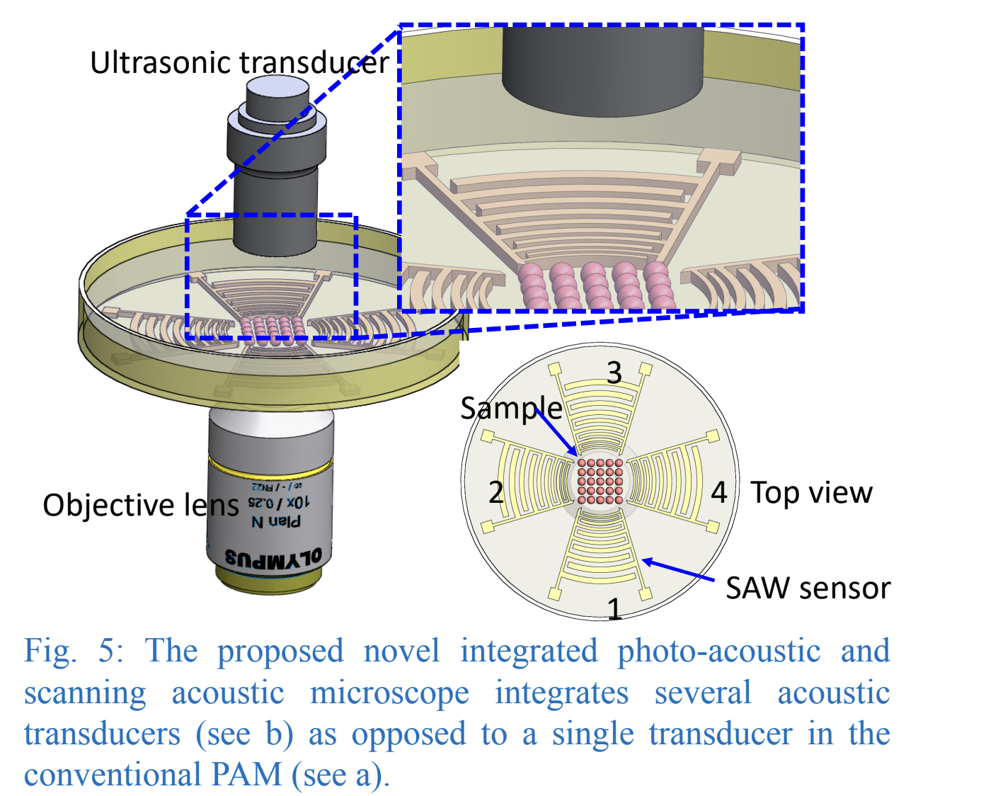
The use of multiple transducers will significantly increase the net angular coverage of the acoustic waves, and thereby the lateral resolution. Acoustic imaging methods used for label-free imaging, are generally absence of toxicity and much more penetrable than optical methods and would therefore give important complimentary information for thicker tissue samples with the possibility to do extract 3D information about tissue elasticity. The use of multiple high frequency sensors for this type of imaging are quite unexplored and would therefore have a high potential for commercialization.